Targeting Cellular Senescence and Skin Aging with Skin Care
What are the best skin care ingredients to reduce cellular senescence in aged skin? Keep reading if you want me to explain what cellular senescence is and why it is important when you want to treat wrinkles on your skin and prevent skin aging.
One of the biggest reasons your skin ages is due to inflammatory factors secreted from senescent cells. This aging process is called inflammaging. What are senescent cells and can ingredients in skin care get rid of these cells that cause skin aging?
Cellular Senescence and Skin Aging
How to get rid of senescent cells and rejuvenate skin?
Preventing cellular senescence is important! Using ingredients that protect skin from pollution and sun radiation and use of sunscreen can help. Anti-inflammatory ingredients and antioxidant ingredients can help prevent senescent cells. Other ways to decrease senescent cells in skin is to stimulate autophagy or activate sirtuin (SIRT-1). (13) SIRT-1 is activated by caloric restriction and by the ingredient resveratrol.
These antiaging skin care ingredients help increase autophagy or decrease cellular senescence
- Aquatide™ , also known as Heptasodium hexacarboxymethyl dipeptide-12
- Crepidiastrum Denticulatum Extract
- Exosomes
- Melatonin (14)
- Pollux CD™ also known as Crepidiastrum Denticulatum Extract)
- Resveratrol (13)
- Saururus Chinensis (15)
- Ulmus Davidiana (16)
Skincare Products that reduce senescent cells and rejuvenate skin
Plated SkinScience Intense Serum has exosomes. Studies have shown that the number of senescent cells is reduced after using this serum. (refs 16, 17)
Cell phases
You need to understand about the different phases in a cell's life cycle so you can understand what cellular senescence is and why it is bad for skin.
Skin cells called keratinocyte and fibroblasts go through five different phases:
Stem (Stem cells)- Think of these as embryo cells that will change significantly as they mature.
Proliferation (Proliferating cells)- These are cells that are dividing and "having babies" and making new cells.
Differentiation (Differentiated Cells)- These are cells that are changing into a specialized cell type.
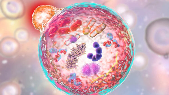
Senescence (Senescent Cells)- These are cells that are not yet dead but are injured and do not function properly.
Apoptosis (Apoptotic Cells)- These are cell going thru programmed cell death. This is nature's way of preventing cancer by getting rid of damaged cells.
The difference between apoptotic cells and senescent cells is that apoptotic cells are not viable and are eliminated while senescent cells, even though they have gone into cell cycle arrest, stay functional and are not eliminated from the skin. We know that an increased number of senescent cells correlates with increased skin aging.
What are Senescent cells?
Senescent cells have lost the ability to proliferate and function properly but have not undergone apoptosis. (1, 2) The lysosomes and mitochondria of senescent cells have lost functionality. (3) They produce harmful secretions that lead to inflammation and other problems that speed aging. The presence of senescence cells is associated with increased aging and seems to speed aging.
What causes cellular senescence?
Normal aging causes cellular senescence. Loss of mitochondrial autophagy also induces senescence as does activation of the TP53 gene, inactivity of SIRT-1, and short telomeres. NRF2 is a key regulator of the skin’s antioxidant defense system, which controls the transcription of genes encoding ROS-detoxifying enzymes and various other antioxidant proteins. (4) Activation of NRF2 induces cellular senescence via direct targeting of certain ECM genes.
Senescent Cells and Skin Aging
Senescent cells are increased in aged skin (3) due to age-related decline of senescent cell removal systems such as the immune system (5) and the autophagy-lysosomal pathway. (7)
You need to understand a bit about what causes skin aging to understand the bad effects of senescent cells.
You can learn about the science of skin aging here.
Senescent cells cause damage and aging of the skin because they because they communicate with nearby cells sending inflammatory cell signals via:
- Matrix metalloproteinases (MMPs)
- Growth factors
- Cytokines
- Chemokines
- Matrix-modeling enzymes
- Lipids
- Extracellular vesicles (EVs).
EVs are lipid bilayer-lined vesicles that can transport functional RNA and microRNA and other modes of communication between cells. (8)
Senescent cells cause a prolonged state of inflammation, known as “inflammaging” (10) which is detrimental to the skin’s appearance. Senescent human fibroblasts secrete proinflammatory cytokines, MMPs and release ROS (11,12) resulting in degradation of the surrounding extracellular matrix (ECM). Loss of the ECM leads to fibroblast compaction and reduced DNA synthesis which disrupts normal cell function and leads to thin, fragile wrinkled skin.
New information is coming out about cellular senescence and what cosmeceutical ingredients are best to treat it. Join us on You Tube or social media to stay current. This is the long interview where Dr. Baumann and Dr. Wyles discuss cellular senescence.
Wondering which skincare products are best to treat your aging skin? Take the skincare routine quiz to build a custom skincare routine from many brands.

Best References and Scientific Publications on Cellular Senescence in Skin
- L. Baumann, Intrinsic Aging in Baumann's Cosmetic Dermatology (McGraw Hill 2022)
- Papadopoulou, A., Kanioura, A., Petrou, P. S., Argitis, P., Kakabakos, S. E., & Kletsas, D. (2020). Reacquisition of a spindle cell shape does not lead to the restoration of a youthful state in senescent human skin fibroblasts. Biogerontology, 1-14.
- Lopez-Otin, C., Blasco, M.A., Partridge, L., Serrano, M., Kroemer, G., 2013. The hallmarks of aging. Cell 153, 1194–1217.
- Hiebert, P., Wietecha, M. S., Cangkrama, M., Haertel, E., Mavrogonatou, E., Stumpe, M., ... & Brinckmann, J. (2018). Nrf2-mediated fibroblast reprogramming drives cellular senescence by targeting the matrisome. Developmental cell, 46(2), 145-161.
- F. Rodier, J. Campisi Four faces of cellular senescence J Cell Biol, 192 (2011), pp. 547-556
- Yoon, J. E., Kim, Y., Kwon, S., Kim, M., Kim, Y. H., Kim, J. H., ... & Kang, H. Y. (2018). Senescent fibroblasts drive ageing pigmentation:? A potential therapeutic target for senile lentigo. Theranostics, 8(17), 4620.
- Dutta, R. Calvani, R. Bernabei, C. Leeuwenburgh, E. Marzetti Contribution of impaired mitochondrial autophagy to cardiac aging: mechanisms and therapeutic opportunities Circ Res, 110 (2012), pp. 1125.
- Terlecki-Zaniewicz, L., Pils, V., Bobbili, M. R., Lämmermann, I., Perrotta, I., Grillenberger, T., ... & Schwingenschuh, S. (2019). Extracellular vesicles in human skin: cross-talk from senescent fibroblasts to keratinocytes by miRNAs. Journal of Investigative Dermatology, 139(12), 2425-2436.
- Campisi, F. d'Adda di FagagnaCellular senescence: when bad things happen to good cells Nat Rev Mol Cell Biol, 8 (2007), pp. 729-740
- Franceschi, J. CampisiChronic inflammation (inflammaging) and its potential contribution to age-associated diseases Gerontol. A Biol. Sci. Med. Sci., 69 (Suppl. 1) (2014), pp. S4-9
- Nelson, J. Wordsworth, C. Wang, D. Jurk, C. Lawless, C. Martin-Ruiz, et al. A senescent cell bystander effect: senescence-induced senescence Aging Cell, 11 (2012), pp. 345-349
- J.F. Passos, G. Saretzki, S. Ahmed, G. Nelson, T. Richter, H. Peters, et al. Mitochondrial dysfunction accounts for the stochastic heterogeneity in telomere-dependent senescence PLoS Biol, 5 (2007), p. e110
- Morselli E, Maiuri MC, Markaki M, Megalou E, Pasparaki A, Palikaras K, Criollo A, Galluzzi L, Malik SA, Vitale I, Michaud M, Madeo F, Tavernarakis N, Kroemer G. Caloric restriction and resveratrol promote longevity through the Sirtuin-1-dependent induction of autophagy. Cell Death Dis. 2010
- Lee, J. H., Moon, J. H., Nazim, U. M., Lee, Y. J., Seol, J. W., Eo, S. K., ... & Park, S. Y. (2016). Melatonin protects skin keratinocyte from hydrogen peroxide-mediated cell death via the SIRT1 pathway. Oncotarget, 7(11), 12075.
Xue-ping, Z. H. E. N. G., Yuan-yuan, F. U., Fan, X. I. A., Qiong, G. U., & Min, L. I. (2019). Discovery and activity studies of ATG4B inhibitors from Saururus chinensis L. Natural Product Research and Development, 31(7), 1252.
Proffer, S. L., Paradise, C. R., DeGrazia, E., Halaas, Y., Durairaj, K. K., Somenek, M., ... & Wyles, S. P. (2022). Efficacy and tolerability of topical platelet exosomes for skin rejuvenation: six-week results. Aesthetic surgery journal, 42(10), 1185-1193.
Dayan, S., Gandhi, N., Wilson, J., Kola, E., Jankov, L. E., Copeland, K., ... & Behfar, A. (2023). Safety and efficacy of human platelet extract in skin recovery after fractional CO2 laser resurfacing of the face: A randomized, controlled, evaluator‐blinded pilot study. Journal of Cosmetic Dermatology, 22(9), 2464-2470.
Plant photo credit: Ptelea, CC BY-SA 4 via Wikicommons


